
We Can Now Watch Your Genome Work In Real Time: Here’s Why That Changes Everything
A landmark review in Nature Reviews Genetics just mapped the living 3D architecture of our DNA. Dr. Andres Zuleta breaks down what CRISPR-Cas live-cell imaging means for your health, and where precision medicine is heading next.
I want you to picture something.
You’re looking at a blueprint for a skyscraper. It’s a flat, two-dimensional sheet of paper, every beam, every electrical wire, every plumbing line laid out in sequence. You can read it. You can understand what should be there. But you can’t see the building actually being built in real time. You can’t watch the workers move, the cranes lift, the wires connect.
For decades, that’s essentially where we were with the human genome.
We cracked the linear sequence of DNA, the “blueprint,” back in the early 2000s. That was extraordinary. But we remained largely blind to what the genome was actually doing inside a living cell at any given moment: which genes were being switched on, which regions of DNA were physically reaching across vast molecular distances to activate one another, how a cell decides to become a liver cell versus a neuron. The genome isn’t a static document. It’s a dynamic, three-dimensional, constantly shifting architecture, and we couldn’t see it in motion.
Until now.
The Problem We Didn’t Know How to Solve
The challenge wasn’t conceptual. Scientists have long theorized that the 3D structure of the genome matters enormously. The problem was physical. There’s a hard limit in conventional light microscopy called the diffraction limit: light waves simply cannot resolve structures smaller than roughly 200 nanometers. Many of the molecular interactions that govern gene activity happen at scales far finer than that, we were trying to watch a conversation happening in a room we couldn’t enter.
What’s more, even if you could image at that scale, doing so in a living cell, without killing it, without disrupting the very processes you’re trying to observe, was another challenge entirely. Most high-resolution imaging techniques require cells to be fixed (essentially killed and frozen in place). Watching life in motion meant you had to let it keep moving.
This is the problem that a landmark review published in Nature Reviews Genetics, authored by Yanyu Zhu, W.E. Moerner, and Lei S. Qi, set out to address. Their work systematically maps the tools, methods, and breakthroughs that have finally — finally — made live-cell genome imaging possible with unprecedented clarity.
What the Researchers Actually Did
This paper is a review, meaning it synthesizes the current state of an entire field rather than reporting a single experiment. Think of it as the scientific community’s best current map of the territory. And what that map reveals is remarkable.
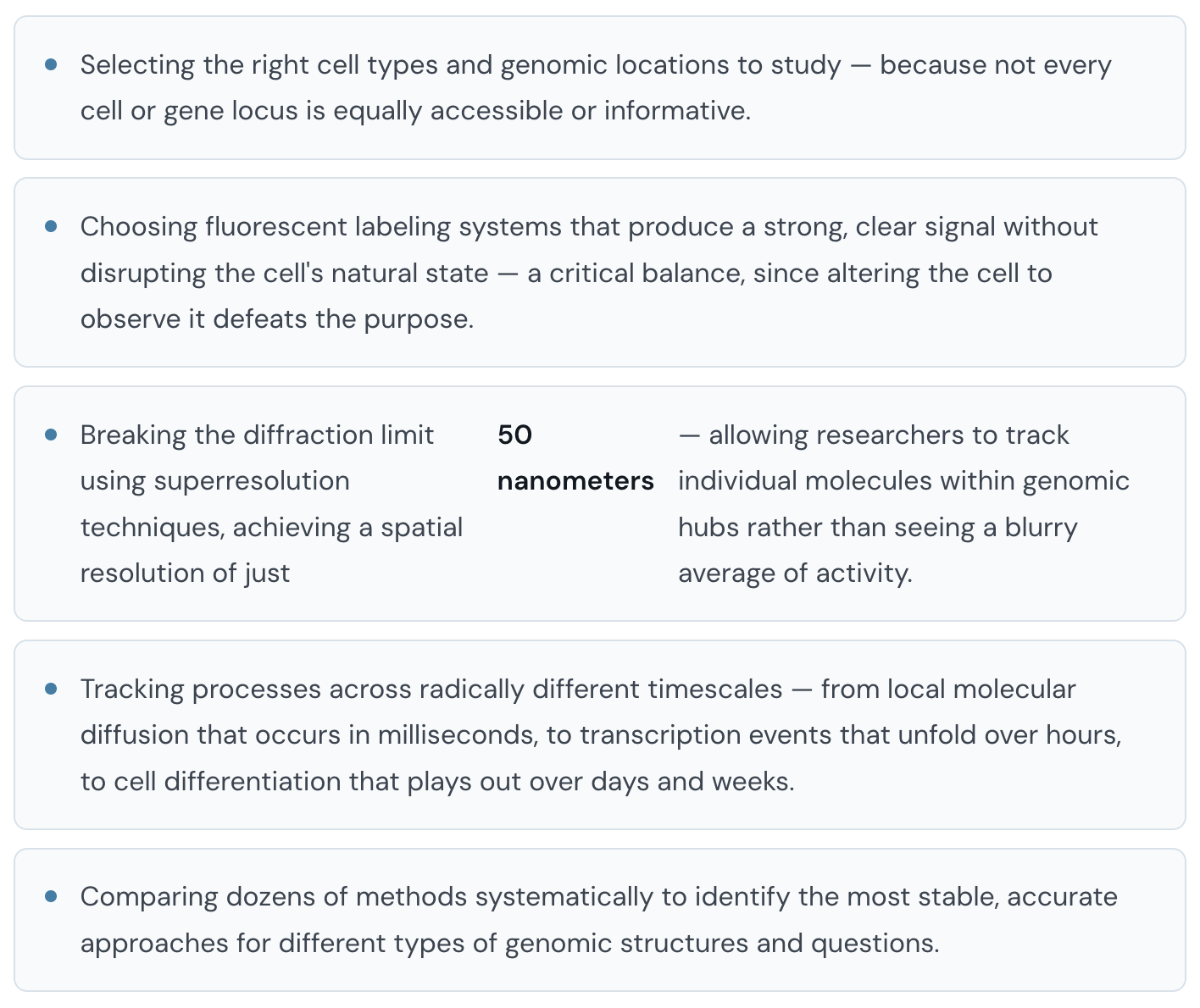
The researchers developed and evaluated a systematic workflow for live-cell genomic imaging. Here’s what that involved, in plain terms:
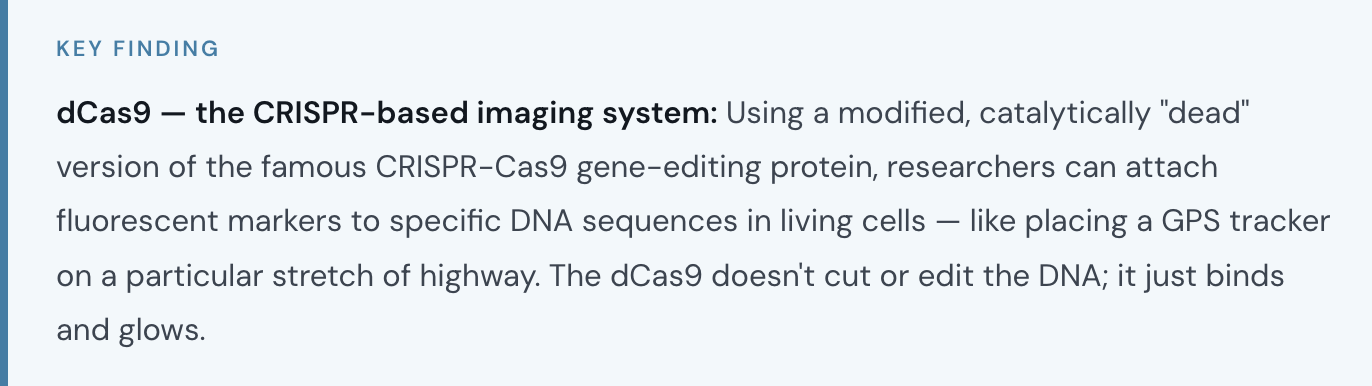
Two specific tools deserve mention because they represent real breakthroughs in the field:
The signal amplification system (dcSCRIBE/SNIPR-based approaches): One persistent problem with fluorescent labeling is photobleaching, the signal fades before you’ve finished watching. These newer amplification systems dramatically extend tracking time without bleaching, enabling long-term observation of genomic events.
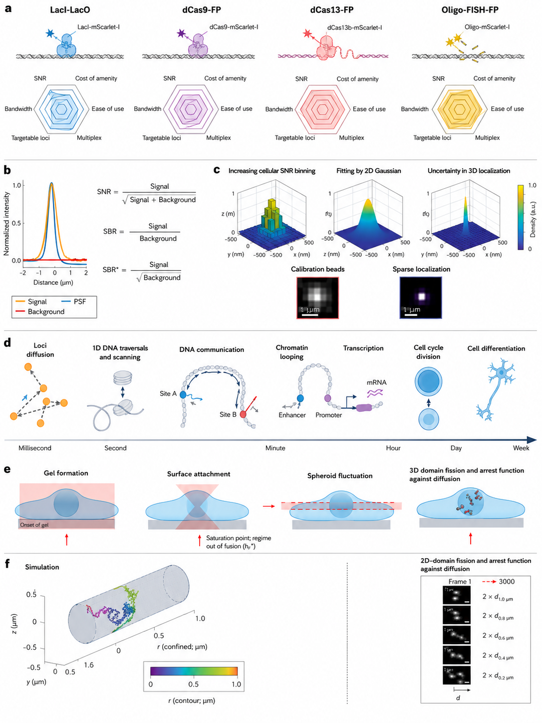
Practical parameters to consider when performing live-cell DNA imaging. Zhu, Moerner & Qi, Nature Reviews Genetics, 2026.
What This Means for Your Health
Here’s where I want to be precise with you, because the science deserves it.
This research is, at its current stage, primarily a tools-and-methods breakthrough. What the Zhu, Moerner, and Qi review establishes is that we now have the capability to watch genome dynamics in living cells with real-time resolution. The downstream applications, for disease diagnosis, for epigenetic medicine, for personalized treatment , are not yet clinical realities. They are, as the researchers put it, doors that have now been opened.
But those doors matter enormously. Think about what it means to observe, in real time, how a gene gets switched on or off by an environmental signal. That’s the core of epigenetics, the study of how our lifestyle, our exposures, our stress levels, our nutrition physically alter which genes are active without changing the underlying DNA sequence itself.

Think about cancer. Many cancers involve misregulated gene expression, genes that should be off are on, or vice versa, often due to disruptions in this 3D chromatin architecture. Being able to observe those disruptions in living cells, in real time, opens the door to earlier detection and more targeted intervention.
Think about aging. Cellular aging involves progressive dysregulation of gene expression, the genome’s architecture becomes noisier, less precise over time. Live-cell imaging may eventually allow us to track these changes longitudinally in individual cells, giving us biological markers of aging that go far deeper than any current biomarker.
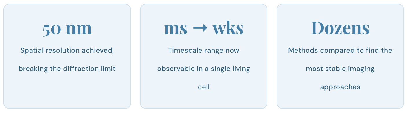
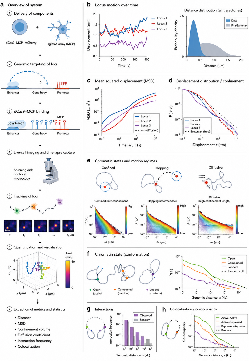
Live-cell imaging data analysis. Zhu, Moerner & Qi, Nature Reviews Genetics, 2026.
The Digital Twin: Where This Is Heading
The researchers point toward something that sounds like science fiction but is grounded in current scientific direction: the biological digital twin.
The concept is this: using real-time genomic imaging combined with AI-driven data analysis, it may eventually be possible to create a data-driven, continuously updated model of your own biological state. Not a generic human model, your model. Your specific 3D genome architecture, your specific epigenetic patterns, your specific cellular dynamics.
With such a model, researchers could theoretically simulate the effect of a treatment, a drug, a dietary intervention, a gene therapy, in a virtual environment before ever applying it to the actual patient. The implications for precision medicine are profound.
I want to be honest: we are years, likely decades, from that being clinical reality. The computational power required, the data integration challenges, the regulatory and ethical frameworks, all of this is still being built. But the imaging tools described in this review are a genuine prerequisite for that future, and those tools now exist.
5 Practical Things You Can Do Starting Now
I know what some of you are thinking: “This is fascinating, Dr. Z, but what does it mean for me right now, today?” Fair point. Here’s what the science, taken as a whole, actually supports as actionable today.
Stay well.
— Dr. Andres Zuleta, MD
Family Medicine Physician | Founder, ThriveMed
For more deep-dives like this one on brain health, longevity, and what the science is actually saying, follow Dr. Zuleta on Medium.
Key References
Zhu Y, Moerner WE, Qi LS. CRISPR-Cas-based live cell imaging of genome dynamics. Nat Rev Genet. 2026. Read →
This article is for educational purposes and is not a substitute for personalized medical advice. If you have specific concerns, please consult a qualified healthcare provider.